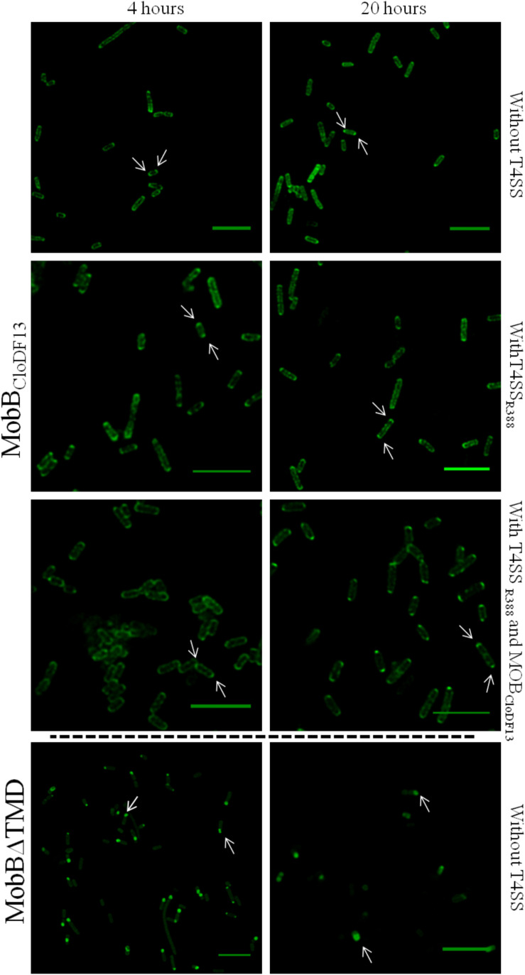
FIGURE 4.
Subcellular location of MobBCloDF13GFP and MobBΔTMDGFP fusion-proteins by confocal fluorescence microscopy. MobBCloDF13GFP and MobBΔTMDGFP proteins were expressed in E. coli BL21C41 (DE3) and E. coli BL21 (DE3) strains, respectively. Subcellular location of these proteins was determined by induction with 1 mM IPTG after 4 (left panels) and 20 h (right panels) at 25°C. Additionally, subcelular location was analyzed in the presence of pSU1456 plasmid, which expresses all R388 conjugative proteins except TrwBR388, or without plasmid pSU1456. MobBCloDF13 was also expressed in the presence of both pSU1456 and pSU4833 that codes for the mobilization proteins of plasmid CloDF13 (MOBCloDF13), except for MobBCloDF13. The images were acquired in a Leica TCS SP5 confocal fluorescence microscope, with a 60× oil immersion objective. Sample excitation was performed with 488 nm wavelength, while fluorescence emission was measured between 505 and 525 nm. The images were analyzed using Huygens and ImageJ software. Arrrowheads indicate the eGFP fluorescence foci at both cell poles (MobBCloDF13) and at a single cell pole (MobBΔTMD). Scale bar: 5 μm.

