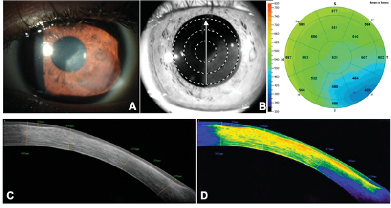
Figure 2.

(A) Slit-lamp photograph of a cornea with inactive nonnecrotizing HSK, showing a diffuse leukoma with no stromal edema. (B) SD-OCT pachymetry showing a thinner cornea (min = 423 µm) in the area of the lesion. (C) A cross-sectional view of the leukoma showing areas of epithelial remodeling and thickening compensation (max = 83 µm) under areas of scarring and stromal compaction (min = 470 µm). (D) Color scale of the lesion showing higher density areas corresponding to fibrosis and scarring on the anterior and medial stroma, fading to yellow and green tones as the lesion becomes less dense.
