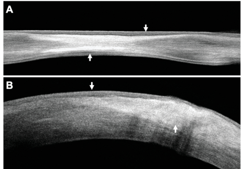
Figure 3.

(A) A cross-sectional image of a cornea with inactive HSK showing epithelial remodeling (top arrow) over a zone of stromal fibrosis and compaction (bottom arrow) with the MCT = 441 µm. (B) A cross-sectional image of a cornea with active HSK showing marked stromal edema (CTL = 641 µm) and an active inflammatory infiltrate (bottom arrow) underlying an area of epithelial edema.
