FIG 7.
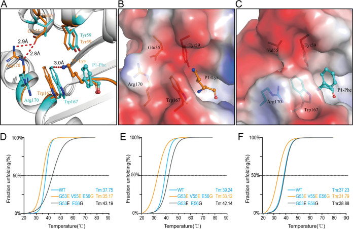
The second opened door at the N terminus of PBG affects peptide assembly. (A) The narrow N terminus of the PBG forces a conformational shift of Trp167. Trp167 of HLA-A*0201 is shown as an orange stick, and Trp167 of RLA-A1 is shown as a cyan stick. The superimposition of RLA-A1 over HLA-A*0201 shows that the conformation of Trp167 predisposes formation of the A pocket of the PBG. The distance of Trp167 in RLA-A1 to P1-Lys in HLA-A*0201 is 3.0 Å, meaning that the conformation shift of Trp167 in RLA-A1 could push the P1 residue of the peptide to the C terminus of the PBG. (B and C) Vacuum electrostatic surface potentials of the N terminus of the PBG of HLA-A*0201 (B) and RLA-A1 (C). Residues Glu55, Tyr59, Trp167, and Arg170 of HLA-A*0201 are shown as orange sticks under the vacuum electrostatic surface. Residues Val55, Tyr59, Trp167, and Arg170 of RLA-A1 are shown as cyan sticks under the vacuum electrostatic surface. P1-Lys of HLA-A*0201 and P1-Phe of RLA-A1 are shown as orange and cyan sticks, respectively. The thermostabilities of RLA-A1 wild type, its substitution mutations at positions 53 and 55, and its substitution mutations at positions 53, 55, and 56 binding to VP60-1 (D), VP60-2 (E), and VP60-10 (F) were tested by circular dichroism spectroscopy.

