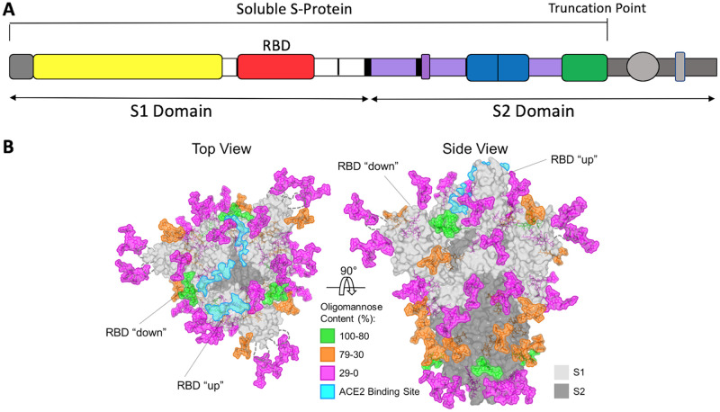
FIG 1.
The SARS-CoV-2 S-protein. (A) Schematic of the S-protein showing the S1 and S2 domains and the RBD. The soluble S-protein ends at the engineered truncation point. The areas colored in gray indicate the transmembrane and intracytoplasmic domains that are present in the full-length S-protein on virions. The most commonly used immunogens are the soluble S-protein, the S1 domain, and the RBD, although some nucleic acid and viral vector constructs are based on the full-length S-protein. (B) Structure-based representation of the S-protein trimer viewed from above and the side, as indicated. The protein surface is in gray, with the ACE2 binding site on the RBD highlighted in aquamarine. On one protomer, the RBD is shown in the “up” position, while on the other two it is in the “down” position, as indicated. Glycans are colored according to the scale, based on their oligomannose content. Adapted from reference 77 under a CC BY 4.0 license.

