
FIG 5.
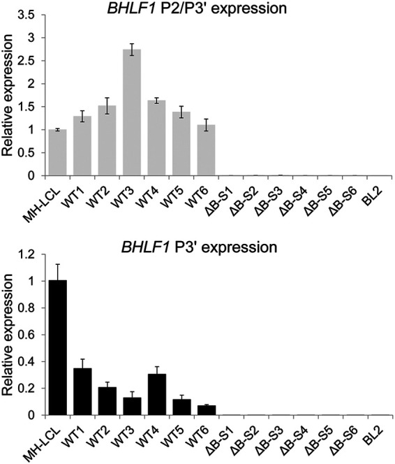
BHLF1 RNA levels within six independently derived BL2 cell lines approximately 1 month after infection with either WT or ΔB-S rEBV were determined by RT-qPCR in triplicate. Error bars indicate the standard deviations. Values are relative to the level of RNA in MH-LCL (LF3−) cells determined using primer sets specific for transcripts initiating from P2 and/or P3′ (P3′-initiating transcripts may overlap those from P2) or at P3′ alone (see diagram in Fig. 2B). RNA from BL2 cells infected with ΔB-S rEBV (BHLF1− LF3+) was included to help exclude the possibility that products amplified with the P2/P3′ and P3′ primer sets had originated from the highly homologous LF3 locus. Comparable results were obtained in separate experiments when amplification was done using a primer set specific for the unique-sequence domain of BHLF1 (data not shown).