FIG 4.
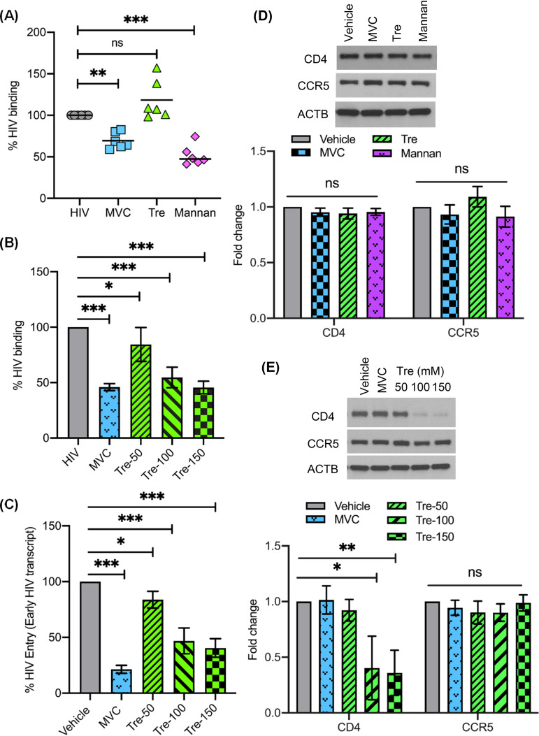
Trehalose inhibits HIV entry into human macrophages. (A) Macrophages pretreated with either 1 μM maraviroc (MVC), 100 mM trehalose (Tre), or 16 mg/ml mannan for 30 min were exposed to HIV for 35 min, after which cells were washed and lysates prepared to detect HIV p24 antigen by ELISA. (B) Macrophages pretreated with either 1 μM maraviroc (MVC) or 50 mM to 150 mM trehalose (Tre-50, Tre-100, Tre-150) for 6 h were exposed to HIV for 35 min, and lysates were assessed for HIV binding by ELISA. (C) Macrophages pretreated with either 1 μM maraviroc (MVC) or 50 mM to 150 mM trehalose (Tre-50, Tre-100, Tre-150) for 6 h were exposed to HIV for 3 h. Following 3 h of incubation, genomic DNA was prepared to measure early virus transcript by qPCR, and results are presented as percent HIV entry compared to vehicle-treated HIV-exposed macrophages. (D) (Top) Representative immunoblot showing expression of CD4, CCR5, and ACTB in cell lysates from vehicle and macrophages treated with MVC (1 μM), trehalose (100 mM), and mannan (16 mg/ml) (30-min treatment). (Bottom) Relative fold change (densitometric analysis) in CD4 and CCR5 protein normalized to ACTB. (E) (Top) Representative immunoblot showing expression of CD4, CCR5, and ACTB in cell lysates from vehicle and MVC-treated (1 μM) and trehalose-treated (50 mM to 150 mM) macrophages (6-h treatment). (Bottom) Relative fold change (densitometric analysis) in CD4 and CCR5 proteins normalized to ACTB. Data are derived from three independent donors and presented as means ± s.e.m. *, P < 0.05; **, P < 0.01; ***, P < 0.001.

