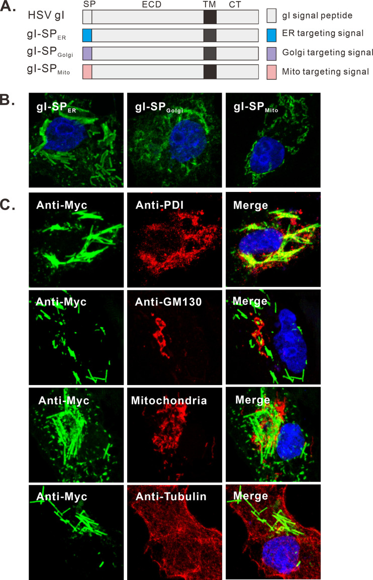
FIG 7.
Analysis of the membrane origin of gI rod-shaped structures. (A) Schematic diagrams of chimeric gI-myc constructs in which the gI signal peptide was replaced with either ER, Golgi, or mitochondrial targeting signal. (B) Vero cells were transfected to express the gI mutants and stained with antibodies to Myc epitope. (C) Vero cells were transfected to express gI-Myc. At 18 to 24 h posttransfection, the cells were fixed, permeabilized, and costained with antibodies to Myc tag (green) and other subcellular markers (red). The cell nuclei were stained with DAPI (blue). The representative images were captured with a Leica confocal microscope and processed by using ImageJ.

