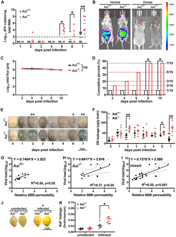
FIG 3.
Effects of Axl on JEV neuroinvasion and blood-brain barrier (BBB) permeability. Four-week-old Axl−/− and Axl+/− mice received an i.p. injection of 104 PFU of JEV (A and E to K) or 5 × 10⁴ PFU of Rluc-JEV for bioluminescent imaging (BLI) (B to D). (A) Shows viral loads in brains; each dot denotes a mouse, and viral loads were compared by two-way ANOVA and multiple t tests. (B) Representative BLI image of Rluc-JEV infection. (C) Quantification of the BLI signal intensity in peritoneal cavity; n = 15 for each group, and the data were compared by two-way ANOVA and multiple t tests. (D) Cumulative percentage of the mice with Rluc-JEV entry into brain; n = 15 for each group, and the data were compared by Fisher’s exact test. (E) Representative image of Evans blue (EB) leakage into brain parenchyma. (F) Quantification of EB leakage into brain parenchyma after JEV infection; n = 5 to 8 for each group, and the data were compared by two-way ANOVA and multiple t tests. (G to I) Correlation analysis of BBB permeability and brain viral load in Axl+/− mice (G), Axl−/− mice (H), and mixed mice (I); each dot denotes a mouse, and the data were analyzed by linear regression model. (J) Representative image of sodium fluorescein (NaF) leakage into brain parenchyma. (K) Quantification of NaF leakage into brain parenchyma under uninfected and JEV-infected (7 dpi) conditions; each dot denotes a mouse, and the data were compared by multiple t tests. All the data are expressed as the means ± SEM; *, P < 0.05; **, P < 0.01; ns, no significance (P > 0.05); each result is the representative of three independent experiments.

