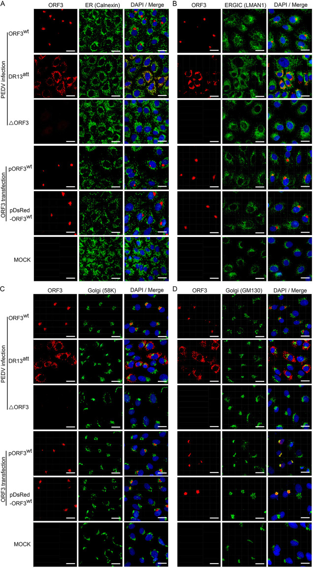
FIG 3.
Intracellular localization of ORF3 protein relative to organelle marker proteins in transfected and infected Vero cells. Vero cells were infected with rDR13att-ORF3wt, DR13att, or rDR13att-ΔORF3 at an MOI of 0.5, transfected with pORF3wt or pDsRed-ORF3wt, or mock infected. After 24 h (infection) or 36 h (transfection) of incubation, the cells were fixed, permeabilized, and processed for IFA using the ORF3 protein antibody P71-3 and appropriate organelle marker monoclonal antibodies, including mouse anti-calnexin (A), mouse anti-LMAN1 (B), mouse anti-58K Golgi protein (C), and mouse anti-Golgi GM130 polyclonal antibody (D) as primary antibodies; Alexa Fluor 647-conjugated goat anti-rabbit IgG (red) and Alexa Fluor 488-conjugated goat anti-mouse IgG (green) were used as secondary antibodies. Separate images showing ORF3 protein localization and organelle marker distribution are presented, as well as mergers of the two. In the merged images the yellow color indicates regions of protein colocalization. Cellular nuclei were stained with DAPI (blue). Scale bar represents 50 μm.

