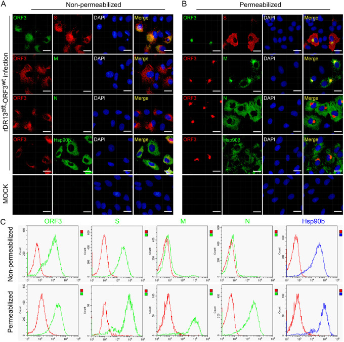
FIG 5.
Detection of the ORF3 protein on the surfaces of PEDV-infected cells. (A and B) Cell surface and intracellular location of PEDV proteins as determined by IFA. Vero cells were infected with rDR13att-ORF3wt at an MOI of 0.5 or mock infected. After 24 h of incubation, the cells were fixed and processed for IFA under both nonpermeabilization (A) and permeabilization (B) conditions. PEDV proteins S, M, and N and ORF3 protein were detected by staining using rabbit anti-S antibody (1:1,000), mouse anti-M antibody (1:100), mouse anti-N antibody (1:100), mouse anti-ORF3 (aa 1 to 71) polyclonal antibody (1:50, top row), or P71-3 (1:50, rows 2 to 4) as primary antibodies. The hsp90β protein, which served as a cell surface marker, was detected by using mouse anti-hsp90β monoclonal antibody (1:100) as a primary antibody. Secondary antibodies were used as for Fig. 4. Cellular nuclei were stained with DAPI (blue). Scale bar represents 50 μm. (C) Cell surface and intracellular location of PEDV proteins as determined by flow cytometry. Cells cultured in 6-well plates were infected with rDR13att-ORF3wt at an MOI of 0.5 or mock infected. After 36 h of incubation, the cells were collected and the cell surface and intracellular proteins were stained under nonpermeabilization (top row) and permeabilization (bottom row) conditions as described for panels A and B, respectively. Flow cytometry was performed as described in Materials and Methods. Data for the PEDV proteins are shown in green and those for hsp90β in blue, while those for an isotype (negative) control are shown in red.

