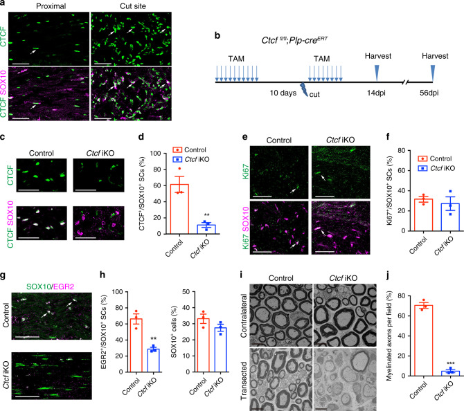
Fig. 5. CTCF is required for SC differentiation during nerve repair.
a Immunolabeling for CTCF and SOX10 in uninjured (proximal) and regenerating region of sciatic nerves of control mice at 14 dpi (n = 3 animals/genotype). Arrows indicate SOX10+/CTCF+ SCs. Scale bars: 50 μm. b A diagram showing the nerve transection scheme. Mice were treated with TAM via i.p. for 10 days, after 10 days, nerves were cut, and mice were then given TAM for 8 days, and nerves were analyzed at dpi 14 and 56. c Immunolabeling for CTCF and SOX10 in regenerating regions of control and Ctcf iKO sciatic nerves 14 dpi (n = 3 animals/genotype). Scale bars: 50 μm. d Proportion of CTCF+ SCs in the regenerating regions of 14 dpi control and Ctcf iKO sciatic nerves (n = 3 animals/genotype). P = 0.0083. e Immunolabeling for Ki67 and SOX10 in the regenerating regions of 28 dpi control and Ctcf iKO sciatic nerves (n = 2 animals/genotype). Arrows indicate representative SOX10+/Ki67+ SCs. Scale bars: 50 μm. f Proportion of Ki67+ SCs in the regenerating regions of 14 dpi control and Ctcf iKO sciatic nerves (n = 3 animals/genotype). P = 0.58. g Immunolabeling of SOX10 and EGR2 in the regenerating regions of 14 dpi control and Ctcf iKO sciatic nerves (n = 3 animals/genotype). Arrows indicate representative SOX10+/EGR2+ SCs. Scale bars: 50 μm. h Proportion of (left) EGR2+ over SOX10+ cells and (right) SOX10+ cells in the regenerating regions of 14 dpi control and Ctcf iKO sciatic nerves (n = 3 animals/genotype). P(left) = 0.005, P(right) = 0.19. i EM images of transverse sections of control and Ctcf iKO 8 weeks after transection (n = 3 animals/genotype). Scale bar: 6 μm. j Proportion of myelinated axons from EM images of control vs. Ctcf iKO 8 weeks after injury (n = 3 animals/genotype). P = 4.26E-05. Data are presented as means ± SEM., **P < 0.01, ***P < 0.001, two-tailed unpaired Student’s t-test. Source data are provided as a Source Data file.

