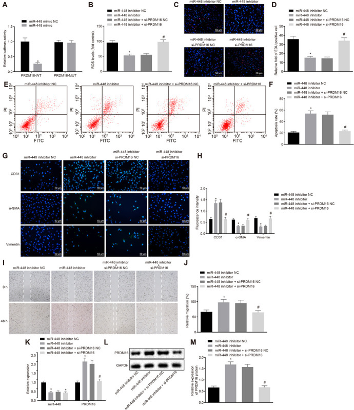
FIGURE 3.

Silencing of miR-448 inhibited the cell proliferation, migration, and invasion while promoting the apoptosis of ASMCs by upregulating the expression of PRDM16. (A) The relative luciferase activity of miR-448 and PRDM16, *p < 0.05 compared with the NC group. (B) ROS levels of cells in each group. (C) Representative images of EdU proliferation staining of cells in each group (×200, scale bar: 50 μm). (D) The relative fold of EdU positive cell in each group. (E) The apoptosis of cells in each group as detected by flow cytometry. (F) The apoptosis rate in each group as detected by flow cytometry. (G) Immunofluorescence of cells in each group (×200, scale bar: 50 μm). (H) The fluorescence intensity of CD31, α-SMA, and vimentin of cells in each group. (I) The cell migration in each group as detected by scratch assay. (J) The relative migration in each group. (K) The relative expression of miR-448 and PRDM16 as detected by RT-qPCR. (L,M) The expression of PRDM16 protein normalized to GAPDH as detected by Western blot analysis. *p < 0.05 compared with ASMCs transduced with miR-448 inhibitor NC, #p < 0.05 compared with ASMCs transduced with miR-448 inhibitor + si-PRDM16 NC. The measurement data were expressed as mean ± standard deviation, and the data of two groups were analyzed by independent-sample t-test. The experiment was repeated three times independently.
