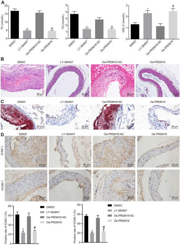
FIGURE 5.

PRDM16 inhibited AS through inactivation of the TGF-β signaling pathway. (A) The content of TC, TG, and HDL-C in peripheral blood in mice in each group as detected by biochemical indicators. (B) The changes of aortic plaques in AS mice as detected by HE staining (×200, scale bar: 50 μm). (C) The number of aortic thrombosis in AS mice as detected by ORO staining (×400, scale bar: 25 μm). (D) Positive rates of AS markers (ICAM-1 and VCAM-1) in the aorta of AS mice as detected by IHC (×400). *p < 0.05 compared with mice treated with DMSO, #p < 0.05 compared with mice treated with oe-PRDM16 NC. The measurement data were expressed as mean ± standard deviation, and analyzed by independent-sample t-test, n = 15.
