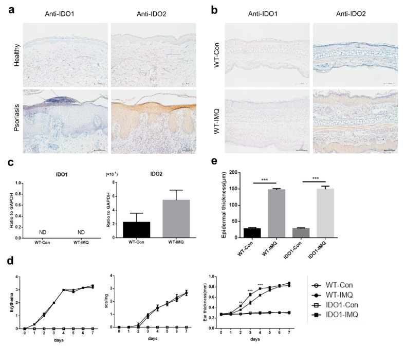
Figure 1.
Psoriasiform lesions in the imiquimod-induced mouse model of psoriasis are associated with IDO2 but not with IDO1. (a) Immunohistochemical staining of the skin from healthy volunteers and patients with psoriasis using antibodies to IDO1 and IDO2. Scale bar = 100 µm. (b) Immunohistochemical staining of the ear from wild-type (WT) mice treated with vehicle or imiquimod (IMQ) for 7 days using antibodies to IDO1 and IDO2. Scale bar = 100 µm. (c) The mRNA levels of IDO1 and IDO2 in the ears of WT mice treated with vehicle or IMQ for 7 days, detected using quantitative real-time PCR. Data are presented as mean ± SEM. (Student’s t-test). Control group, n = 6; IMQ-treated group, n = 9. (d) Erythema, scaling, and thickness of the ear in WT and IDO1 KO mice treated with vehicle or IMQ for 7 days were evaluated daily (0 = none, 1 = slight, 2 = moderate, 3 = marked, 4 = very marked). ** p < 0.01, *** p < 0.001 when comparing IDO1 KO-IMQ with WT-IMQ (two-way ANOVA). Control groups, n = 3; IMQ-treated groups, n = 6. (e) Microscopic evaluation of epidermal hyperplasia of the ear in WT and IDO1 KO mice treated with vehicle or IMQ for 7 days. Data are presented as mean ± SEM. *** p < 0.001 (one-way ANOVA). Control groups, n = 3; IMQ-treated groups, n = 6. ND: not-detected, WT: wild type, IDO: indoleamine 2,3-dioxygenase, Con: control, IMQ: imiquimod, KO: knockout.

