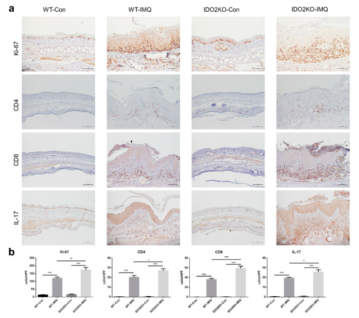
Figure 4.
Keratinocyte proliferation and inflammatory cell infiltration are significantly increased in IDO2 KO mice treated with IMQ. (a) Immunohistochemical staining of the ear of WT and IDO2 KO mice treated with vehicle or IMQ for 7 days using Ki-67, CD4, CD8, and IL-17 antibody. Ki-67 antibody, scale bar = 50 µm; CD4, CD8, and IL-17 antibody, scale bar = 100 µm. (b) The number of Ki67+ cells in the epidermis and infiltrating CD4+, CD8+, and IL-17+ lymphocytes in the dermis per high power field. Data are presented as mean ± SEM. * p < 0.05, ** p < 0.01, *** p < 0.001 (one-way ANOVA). Control groups, n = 6–7; IMQ-treated groups, n = 9. WT: wild type, IDO: indoleamine 2,3-dioxygenase, Con: control, IMQ: imiquimod, KO: knockout.

