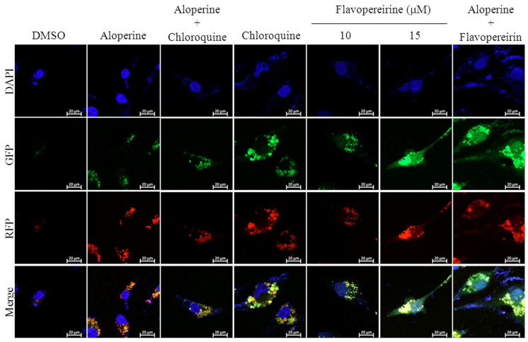
Figure 2.
Flavopereirine inhibited autophagic flux in MDA-MB-231 cells. Representative fluorescent images of autophagosomes and autophagolysosomes in MDA-MB-231 cells were transduced with tandem RFP-GFP-LC3B and then independently treated with aloperine (100 μM), chloroquine (25 μM), flavopereirine, or a combination of the two drugs. In the merged images, autophagosomes are presented as yellow or orange puncta (RFP-GFP-LC3B), whereas red puncta (RFP-LC3B) indicate autophagolysosomes because acidification abolishes the green fluorescence. Images were obtained using a 63× oil immersion objective.

