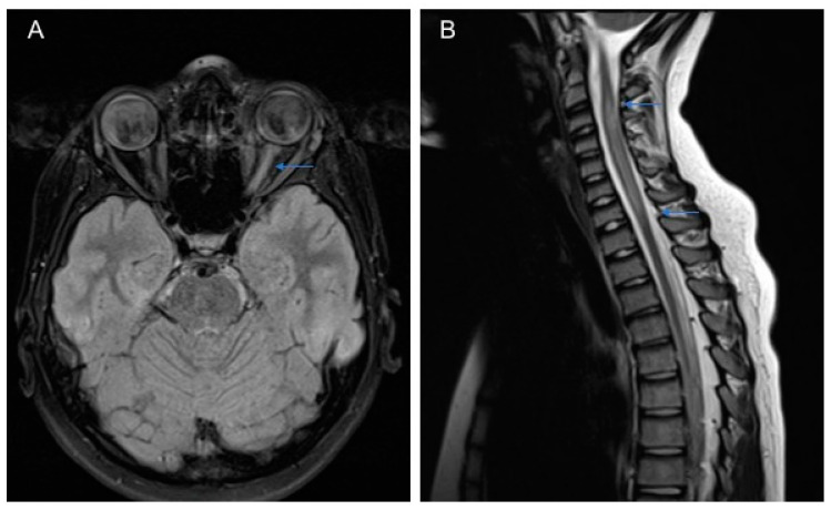
Figure 1.
MRI of a 11-year-old patient with Neuromyelitis optica (NMO): (A) the brain axial Flair-T2 weighted image shows hyperintensity of the left optic nerve and (B) the spinal T2 weighted image shows cervical hyperintense lesions extending longitudinally from C3 to C7 (longitudinally extensive transverse myelitis (LETM).

