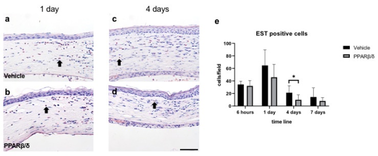
Figure 2.
EST staining. EST staining was performed to examine neutrophil infiltration. Vehicle group (a,c) and PPARβ/δ group (b,d) in the corneal limbus on day 1 and day 4 after injury (black arrows: EST-positive cells). Bar, 50 μm. Fewer EST-positive neutrophils were present in the PPARβ/δ group than in the vehicle group (e). Data are presented as mean ± SD (n = 8) * p < 0.05.

