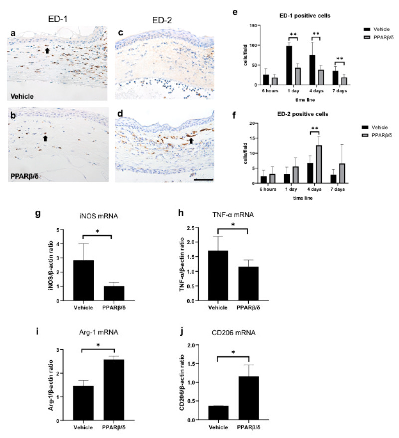
Figure 6.
Immunostaining for ED-1 and ED-2. Pan-macrophages are stained with ED-1, and M2 macrophages are stained with ED-2. ED-1 immunostaining (a: vehicle group, b: PPARβ/δ group) in the corneal limbus on day 1. ED-2 immunostaining (c: vehicle group, d: PPARβ/δ group) in the corneal limbus on day 4. Black arrows: immunostained cells. Bar, 50 μm. ED-1-positive cells were lower on days 1, 4, and 7. On the other hand, ED-2-positive cells were higher on day 4 in the PPARβ/δ group versus the vehicle group (e, f). The mRNA levels of the indicated M1 (iNOS and TNF-α) (g, h) and M2 (Arg-1, and CD206) (I, j) at 6 h. The M1 markers were lower and the M2 markers were higher in the PPARβ/δ group as compared to the vehicle group. Data are presented as mean ± SD (n = 8) ** p < 0.01 or * p < 0.05

