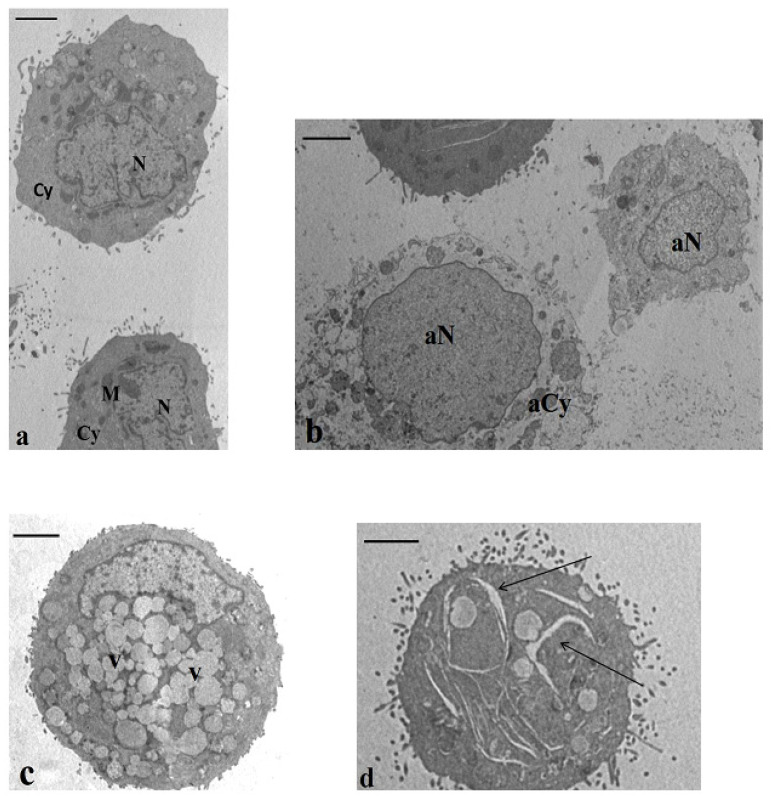
Figure 5.
Transmission electron microscopy (TEM) sections of cultured HT-29 cells. (a) Baseline conditions. The cells showed normal nuclei (N), regular chromatin texture, and cytoplasm (Cy) containing the typical organelles’ mitochondria (M). Bar 3.5 μm. (b–d) Incubation with CB83, THC, and CBD. In all samples, a high percentage of cells with necrotic and apoptotic features was described. In (b), a necrotic cell is evidenced in an altered chromatin texture (aN) and a cytoplasm devoid of organelles (aCy) and, in (c), is represented in an apoptotic cell with a cytoplasm rich in vacuoles (V). The cell showed in (d) highlights enlargements in the cytoplasm (arrows). This feature was detected in a percentage of 8–12% in all treated samples. (b,d) Bar 3.5 μm. (c) Bar 3 μm.

