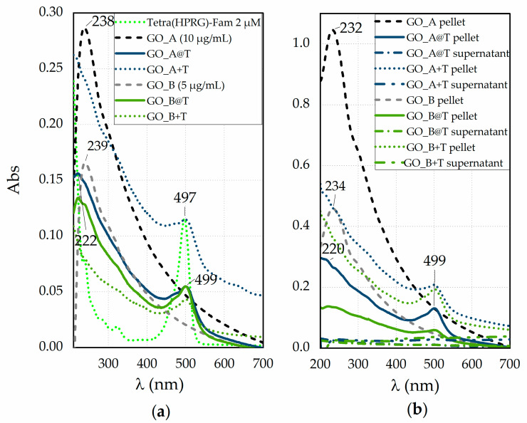
Figure 1.
(a) UV-visible spectra in phosphate buffer saline solution (PBS, pH = 7.4) of GO_A and GO_B 100× diluted dispersions, both before (dashed lines for GO_A, in black, and GO_B, in grey, respectively) and after 2 h of incubation with the peptide (solid line for GO_A@T, in dark green, and GO_B@T, in light green, respectively). The reference spectra of Tetra(HPRG)-Fam (green, 100× diluted solution of that used for the incubation of GO_A and GO_B samples) and the mixtures immediately the mixing (GO_A+T, in dark green, and GO_B+T, in light green, respectively) are shown for comparison in dot line. (b) UV-visible spectra in PBS of the pellets for hybrid GO_A@T and GO_B@T samples (solid lines), the GO_A+T and GO_B+T mixtures (dot lines) and, for comparison, of bare GO_A and GO_B pellets (dashed lines) after two centrifugation (2700 RCF, 2 min, RT) and washing steps. The spectra of supernatants (dashed dot lines) of GO@T samples collected after the first washing step are included.

