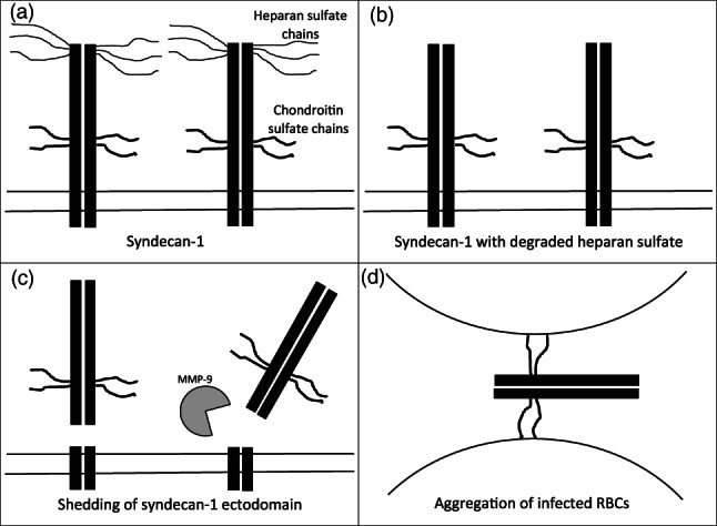
Figure 2.

Schematic representation of the proposed sequential changes in syndecan‐1 in placental malaria. (a) A depiction of intact syndecan‐1 embedded in the plasma membrane of the syncytiotrophoblast showing the heparan sulfate chains on the ectodomain distal from the cell surface and the chondroitin sulfate chains on the ectodomain proximal to the cell surface. (b) Syndecan‐1 after the heparan sulfate chains have been removed by heparanase. (c) The removal of the heparan sulfate chains has cleared the way for MMP‐9 to access the ectodomain cleavage site near the surface of the syncytiotrophoblast. (d) The cleaved syndecan‐1 ectodomain with chondroitin sulfate chains has been released, allowing it to bind to multiple infected RBCs. The depictions of syndecan‐1 shown here were influenced by related models shown in Elenius and Jalkanen (1994), Bernfield et al. (1999), Pries et al. (2000), Manon‐Jensen et al. (2010), Roper et al. (2012), and Itoh et al. (2015). HS = heparan sulfate; infected RBCs = infected red blood cells; MMP‐9 = matrix metalloproteinase 9
