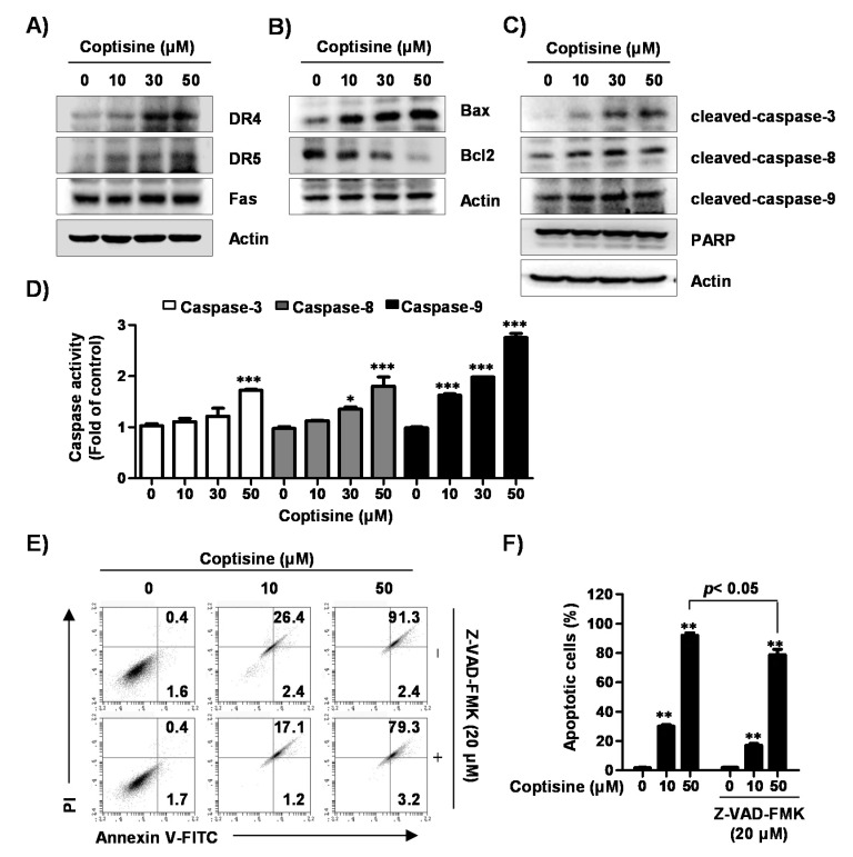
Figure 3.
Effects of coptisine on extrinsic and intrinsic apoptotic pathways in Hep3B cells. (A–C) Hep3B cells were treated with coptisine for 24 h, cells were harvested for obtain total cell lysates. Cell lysates was separated by sodium dodecyl sulfate polyacrylamide gel (SDS-PAGE) and transferred to polymerization of vinylidene difluoride (PVDF) membranes. The membranes were blocked, probed with indicated antibodies, and incubated with secondary antibodies for detection; (D) After cells were treated with the indicated concentration of coptisine for 24 h, cells were harvested, and incubated with each substrate. The activities of caspases were detected by a microplate reader. Data are presented as the mean ± SD of three independent experiments. * p < 0.05 and *** p < 0.001 vs. control; (E) After cells were treated with benzyloxycarbonyl-Val-Ala-Asp (OMe) fluoromethylketone (Z-VAD-FMK), a pan caspase inhibitor, for 1 h, cells were treated with coptisine for 24 h. The cells were collected, stained with annexin V-FITC and PI, and analysis by flow cytometry; (F) Shows the percentage of annexin V+ cells. The results are presented as the mean ± standard deviation (SD) of three independent experiments. ** p < 0.01 vs. control.

