Figure 2.
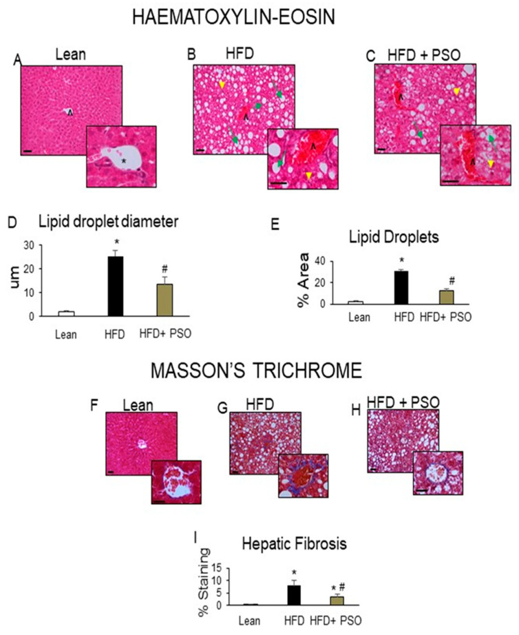
Histological analysis of livers. In lean mice, (A–C) Representative Haematoxylin-eosin (H&E) staining and (D,E) quantitation showed (A) a normal hepatic parenchyma with radially arranged rays of hepatocytes, regularly directed from the central vein of each lobule towards its periphery. (B) HFD-treated mice exhibited significant morphological alterations with lipid droplets deposition and inflammatory cells infiltration. (C) Hepatic parenchymal alterations in the HFD group were decreased in HF animals treated with PSO. (D) Quantitation of lipid droplet diameter. (E) Quantitation of the lipid droplet area. (F) Hepatic fibrosis by Masson’s staining in lean mice that exhibited very weak parenchymal fibrosis; (G) HFD mice demonstrating strong perisinusoidal collagen deposition; (H) HFD + PSO-treated mice demonstrating reduced perisinusoidal collagen deposition; (I) quantitation of collagen staining. * indicates a centrolobular vein. Green arrows indicate lipid droplets and yellow arrowheads indicate areas of lobular inflammatory loci. Bar = 50 µm. * p < 0.05 from corresponding value in lean mice. # p < 0.05 from corresponding value in HFD mice. n = 6/group.

