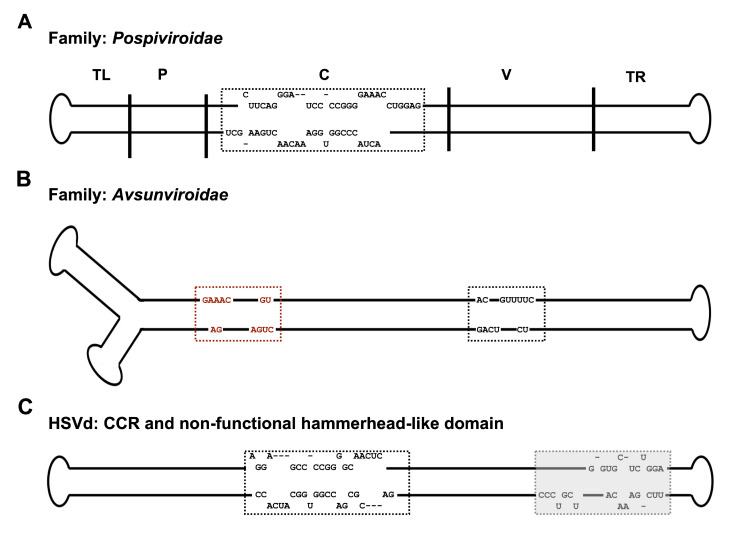
Figure 2.
Primary and secondary structural features used for viroid classification. Schematic representations of (A) The rod-like secondary structure of potato spindle tuber viroid (PSTVd). The five functional domains are shown on the secondary structure of PSTVd: The Terminal left (TL), Pathogenicity (P), Central (C), Variable (V), and Terminal right (TR) domains are delimited by the vertical solid lines and are named accordingly. The sequence of the central conserved region (CCR), which is the characteristic feature of the members of the family Pospiviroidae, is indicated within the box. (B) The branched secondary structure of avocado sunblotch viroid (ASBVd). The conserved nucleotides of the hammerhead’s catalytic core, the characteristic feature of the members of the family Avsunviroidae, are boxed. The sequences within the red and black boxes denote the hammerhead self-cleaving motifs formed in the viroid’s (+) and (−) strands, respectively. (C) The rod-like secondary structure of hop stunt viroid (HSVd) showing both the CCR (black color boxed) and a non-functional hammerhead-like domain (shadowed box), respectively.

