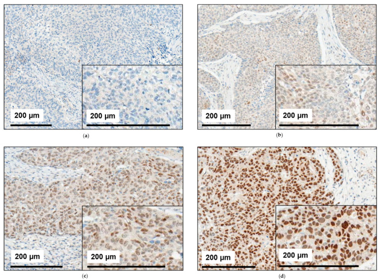
Figure 1.
Staining patterns for NR2F6 in HNSCC. The HNSCC tissue from our cohort was immunohistochemically assessed for NR2F6 expression. (a) Some tumors showed no NR2F6 expression. In other tumors, the mean NR2F6 expression was (b) low, (c) medium, or (d) high. Some HNSCC also showed an inhomogeneous NR2F6 expression with (b,c) positive and negative cells.

