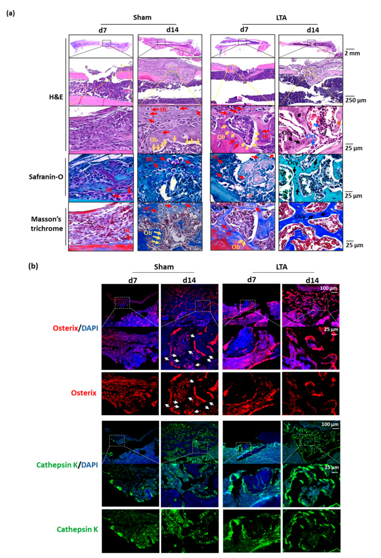
Figure 2.
Lipoteichoic acid (LTA) exerts beneficial effects on bone morphology, osterix expression and cathepsin K expression in mice with femoral bone defects. (a) Hematoxylin and eosin (H&E), safranin-O and Masson’s trichrome staining revealed the presence of multiple cell types during the bone-regeneration process in the longitudinal histological sections. The bone bridge, hypertrophic chondrocytes (HCs), and osteoblasts (Obs) were observed on Day 14 after femoral bone defects were introduced in the sham control group. The bone bridge, HCs and Obs appeared earlier in the LTA group, on Day 7 after femoral defects were introduced. An extensive network of primary bone (osteocytes, Ocys) and osteoclasts (Ocls) had formed and woven bone was observed on Day 14 following the introduction of femoral defect in the LTA group. (b) Immunofluorescence was used to detect osterix (an Ob marker) and cathepsin K (an Ocl marker). Intense osterix and cathepsin K signals were observed surrounding the trabecular bones in the LTA-treated group, whereas the signals for these two proteins were diffusely distributed in the PBS group. The white arrows indicate that fewer newly formed bones were present in the sham group, whereas vigorous bone-formation activity was observed in the LTA group.

