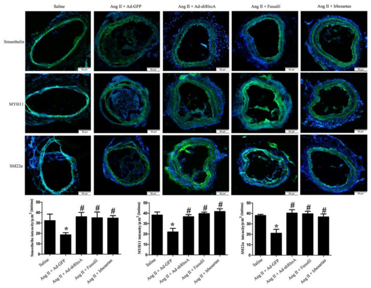
Figure 10.
Immunofluorescence staining of MYH11, SM22α, and smoothelin (green). Nuclei were stained with DAPI (blue). Mice were treated the same way as in tablee 7. Histograms show the fluorescence intensity of the staining in the intima. * p < 0.05 vs. the saline-infused group; # p < 0.05 vs. the model group (Ang II infusion + Ad-GFP virus) (n = 10). The scar bar is 50 μm.

