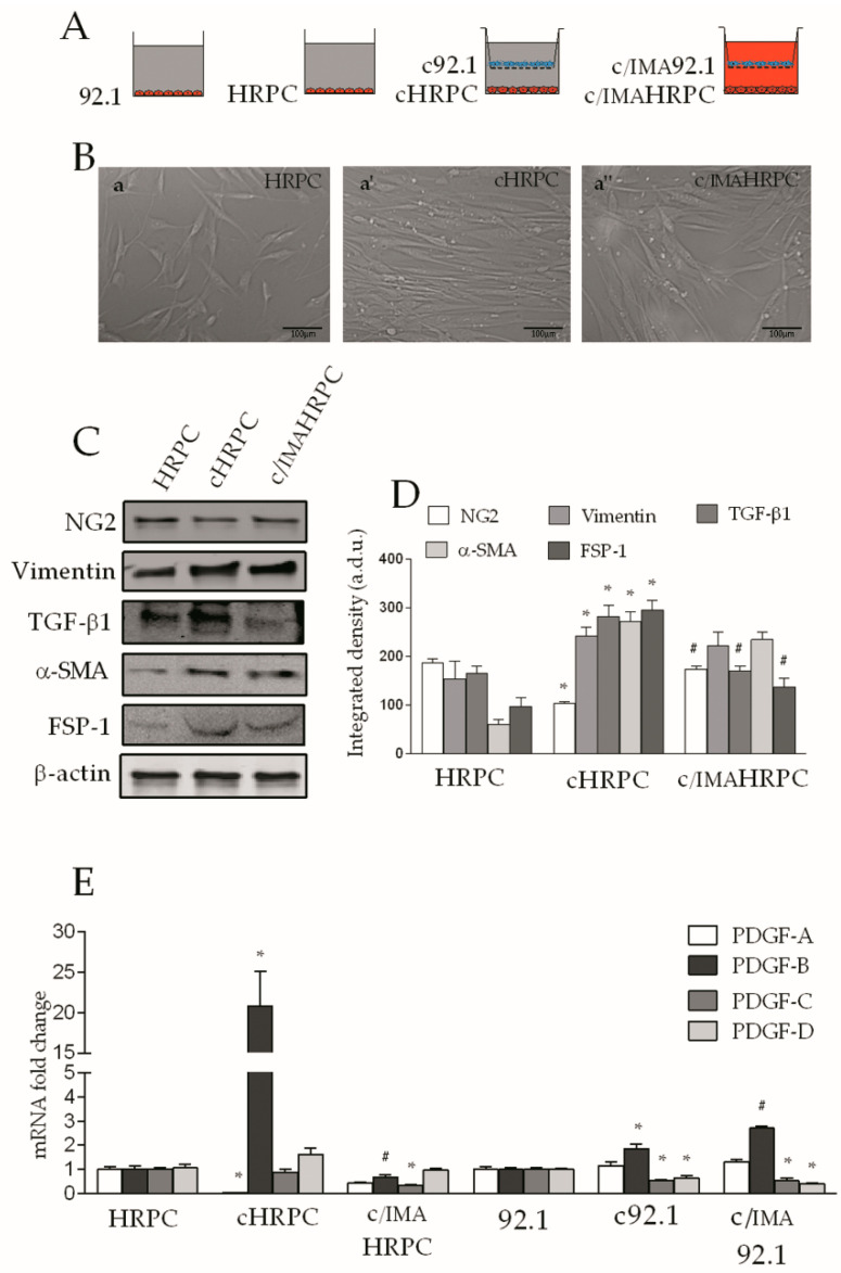
Figure 1.
Imatinib prevents marker expression changes in human retinal pericytes (HRPC) induced by the interaction with 92.1 uveal melanoma (UM) cells. Human retinal pericytes were grown alone (control HRPC) or were conditioned in the presence of 92.1UM (cHRPC) with or without 5 μM of imatinib (c/IMAHRPC) for 6 days (A,B): representative images of HRPC (a), cHRPC (a’), and c/IMAHRPC (a’’) after 6 days of conditioning. (C): Western blot analysis of NG2, vimentin, TGF-β1, α-SMA, and FSP-1 proteins in control pericytes (HRPC), cHRPC, and c/IMAHRPC. β-actin detection indicates the same loading of 30 μg of protein in each lane. (D): immunoblot quantification of through densitometric analysis of each band (in arbitrary densitometry units, a.d.u.), carried out with the Image J program. (E): quantification of PDGF-A, PDGF-B, PDGF-C, and PDGF-D mRNA expression levels by qRT-PCR in both HRPC and 92.1UM grown alone and in coculture with or without 5 μM of imatinib. Bars represents the means ± SEM from three independent experiments. * p < 0.05 vs. control (HRPC or 92.1UM); # p <0.05 vs. coculture without imatinib. One-way ANOVA, followed by Tukey’s test.

