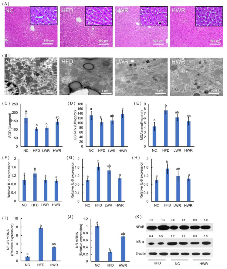
Figure 2.
Wild rice prevents HFD-induced adipocyte hypertrophy, hepatic steatosis, and systemic inflammation. (A) Hematoxylin and eosin (HE)-stained liver sections. (B) Ultrastructural changes in the liver were detected via TEM. The (C) liver superoxide dismutase (SOD), (D) GSH-Px, and (E) malondialdehyde (MDA) were measured using commercial assay kits. The (F) interleukin (IL)-1, (G) IL-6, and (H) IL-8 concentrations were measured using ELISA. Gene expression levels of (I) NFκB and (J) IκB-α in the liver were measured by qRT-PCR. (K) Liver NFκB and IκB-α protein production was examined by Western blot, and relative protein levels were normalized with β-actin (n = 3). (C–J) a,ab,b: The values of each group with the same letters are not significantly different in the analysis of variance followed by Tukey’s post-hoc test.

