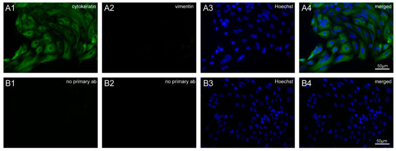
Figure 3.
Immunofluorescence staining of isolated bovine oviductal epithelial cells (BOECs). (A1) Cells incubated with anti-cytokeratin antibody (green) showing positive staining. (A2) No signal was found on the incubation of cells with the anti-vimentin antibody (red). (A3) Staining of nuclei with Hoechst (blue). (A4) Overlay. (B1,B2) Negative control staining without primary antibodies. (B3) Hoechst staining and (B4) overlay of control cells. Images were captured with a Leica DM5500 B microscope equipped with Leica DFC310 camera and processed with ImageJ.

