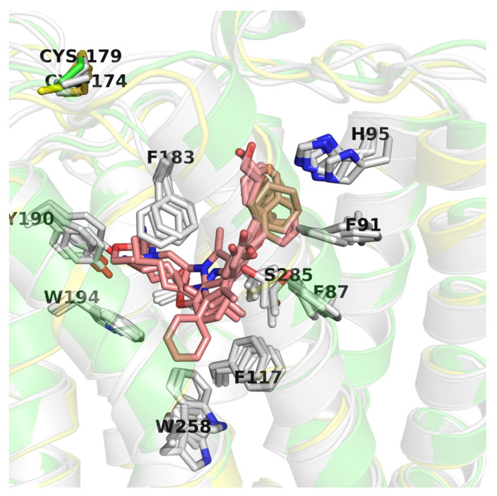
Figure 7.
Comparison of CB2 structures available in the Protein Data Bank (PDB). Here, an inactive CB2 conformation (5ZTY) is shown in green and is the best performing in virtual screening (VS), active conformation (6KPF) is shown in yellow, and other CB2 active structures are shown in grey. Residues are labeled according to the 5ZTY numbering.

