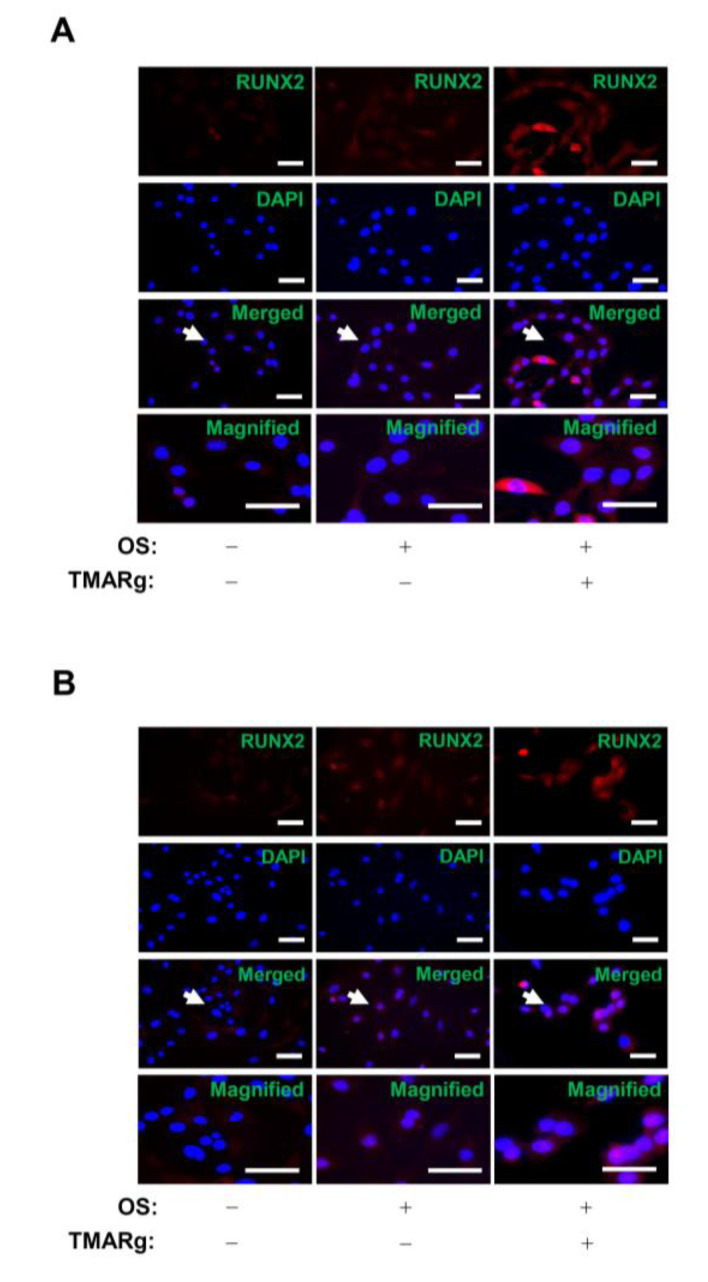
Figure 5.
TMARg accumulates the expression of RUNX2 into the nucleus in osteoblast differentiation. (A,B) After mesenchymal cells (A) and pre-osteoblasts (B) were cultured in OS with TMARg (30 µM) for 24 h, the cells were fixed and permeabilized. RUNX2 was immunostained with rabbit anti-RUNX2 antibody, followed by Alexa-Fluor 568-conjugated secondary antibody (red). Then, the cells were counterstained with DAPI (blue). The third panel shows the merged images of the first and second panels. The bottom panels show the magnifications of the merged images. The arrow indicates the magnified region. Scale bar: 50 µm. The data are representative of the results of three independent experiments.

