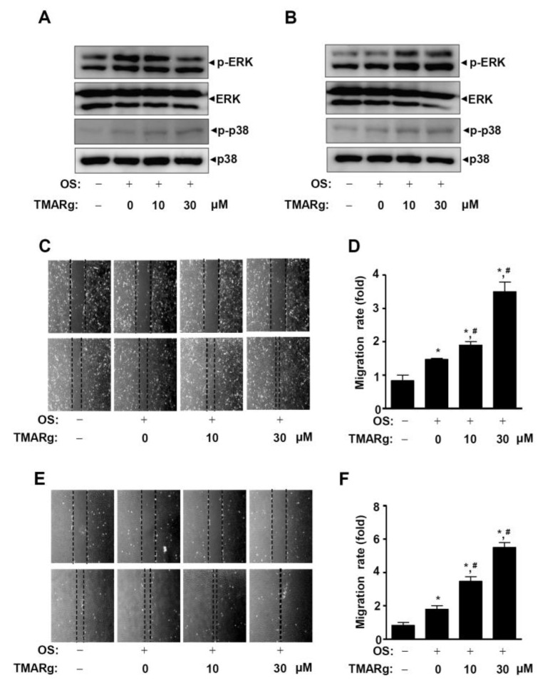
Figure 6.
TMARg activates MAPKs signaling and promotes the cell migration rate in osteoblast differentiation. (A,B) After mesenchymal cells (A) and pre-osteoblasts (B) were cultured in OS with TMARg (10 and 30 µM) for 24 h, phospho-ERK1/2, ERK1/2, phospho-p38, and p38 were analyzed using Western blot analysis. (C–F) Cell migration in mesenchymal cells (C,D) and pre-osteoblasts (E,F) was observed under a phase contrast microscope (C,E), and the cell migration rate (fold) was measured by the area enclosing the spreading cell population and expressed as a bar graph normalized to the control (D,F). The data are representative of the results of three independent experiments. Data represent the means ± SEMs of experiments. *, p < 0.05 was considered significantly different, compared to the control. #, p < 0.05 was considered significantly different, compared to the OS.

