
Figure 5.
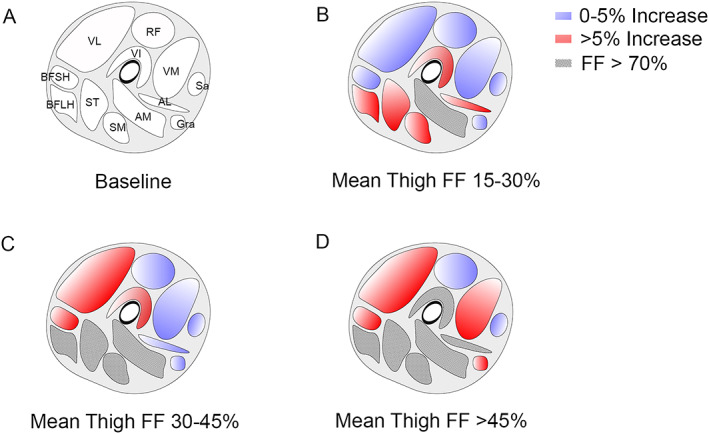
Progression of fat fraction depending on the baseline mean thigh fat fraction. (A) Baseline image showing the localization of thigh muscles (VL: vastus lateralis, RF: rectus Femoris, VM: vastus medialis, VI: vastus intermedius, BFSH: biceps femoris short head, BFLH: biceps femoris long head, ST: semitendinosus, SM: semimembranosus, AM: adductor major, AL: adductor longus, Sa: sartorius, and Gra: gracilis). (B) Increase in fat fraction over a 4 year period in patients with baseline mean thigh fat fraction of 15% to 30%. (C) Increase in fat fraction over a 4 year period in patients with baseline mean thigh fat fraction of 30% to 45%. (D) Increase in fat fraction over a 4 year period in patients with baseline mean thigh fat fraction >45%. FF, fat fraction.