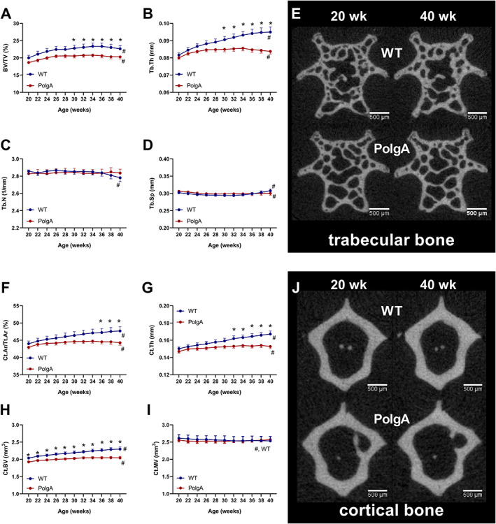
FIGURE 6.

Longitudinal monitoring of trabecular and cortical bone morphometric parameters over 20 weeks: (A) bone volume fraction (BV/TV), (B) trabecular thickness (Tb. Th), (C) trabecular number (Tb. N), (D) trabecular separation (Tb. Sp), (F) cortical area fraction (Ct.Ar/Tt.Ar), (G) cortical thickness (Ct. Th), (H) cortical bone volume (Ct.BV), and (I) cortical marrow volume (Ct.MV). (E,J) Bone microstructure (cross sections) of representative (median) wild type (WT) (top row) and PolgA (bottom row) mice at 20 (left) and 40 (right) weeks of age. In WT mice, thickening of trabeculae and cortex can be observed, while in PolgA mice, little change can be seen between time points. Data represent mean ± SEM; n = 9 WT and n = 12 PolgA, * P < 0.05 WT (blue line) vs. PolgA (red line); #P < 0.05 over time determined by linear mixed model and Tukey's post‐hoc.
