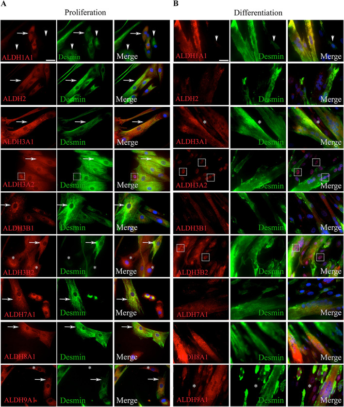
Figure 7.

Immunocytological phenotyping of primary muscle cells in culture. Human cells were extracted from TFL muscles of healthy donors and grown in culture in proliferation medium (two passages, A) or in proliferation (two passages) and then in differentiation medium (4 days, B). Cells were fixed using PFA and permeabilized using saponin; and then incubated with antibodies directed against ALDH isoenzymes, and desmin; and then incubated with labelled secondary antibodies. Nuclei were stained using DAPI (blue). In proliferation (A), the number of cells expressing isoenzymes (red, first column) generally exceeded the number of cells expressing desmin (green, second column) (ALDH3A2, 3B2, 7A1, 8A1, and 9A1, arrows) except for 1A1 where some desmin+ cells were negative for the isoenzyme. ALDH3A2 stained strongly some nuclei. Following differentiation (B), myotubes were generally labelled with a stronger intensity than mononucleated cells (ALDH3A1, 3B1, and 9A1). The intensities of ALDH1A1 and 2 labelling were decreased as compared with cells in proliferation. Several nuclei strongly expressed ALDH3A2, suggesting a translocation from cytoplasm to nucleus. Original magnifications: ×40.
