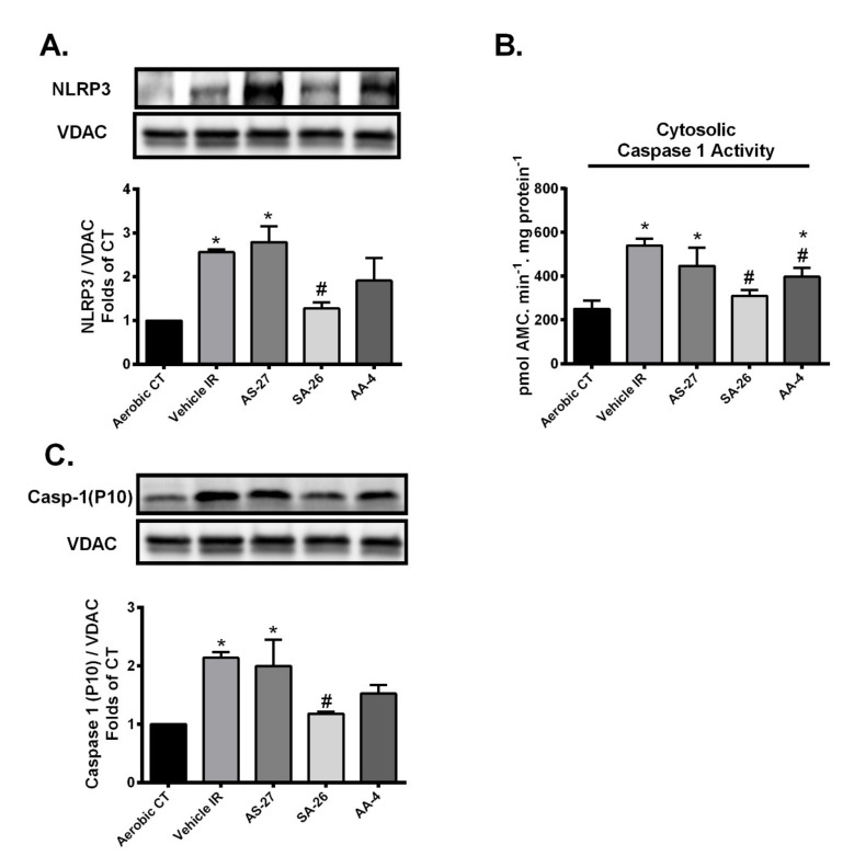
Figure 5.
Perfusion of hearts with SA-26 inhibited the IR-induced activation and translocation of the NLRP3 inflammasome to the mitochondria. (A) Representative immunoblots and densiometric quantification of the mitochondrial expression of NLRP3 protein in mice hearts after 30 min ischemia and 40 min reperfusion. Protein expression was normalized to VDAC loading control. (B) Cardiac caspase-1 enzymatic activity assessed in the cytosolic fraction following 30 min ischemia and 40 min reperfusion. The assay quantitated the fluorescence intensity resulting from the cleavage of the caspase-1 specific fluorogenic substrate Ac-YVAD-AMC by the cytosolic heart homogenates. (C) Representative immunoblots and densiometric quantification of the mitochondrial expression of cleaved caspase-1 (P10) in mice hearts after 30 min ischemia and 40 min reperfusion. Protein expression was normalized to VDAC protein used as a loading control. Values represent mean ± SEM, * p < 0.05 vs. Aerobic CT, # p < 0.05 vs. Vehicle IR (n = 4 –5 per group).

