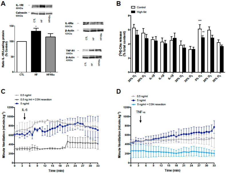
Figure 4.
Effect of hypercaloric diets on the expression of pro-inflammatory cytokines receptors in CB and effect of pro-inflammatory cytokines on ventilation and catecholamine release from the carotid body (CB). (A) Effect of high-fat (HF) for 3 weeks and high-fat high-sucrose (HFHSu) diet for 25 weeks on the expression of the receptors IL-1RI, IL-6Rα, and TNF-R1 in rat CB. (B) Effect of interleukin-1 beta (IL-1β, 40 ng/mL) on the release of catecholamines from the CB in control and HF animals. (C,D) Effect of interleukin-6 (IL-6, 0.5 or 5 ng/mL) and tumor necrosis factor alpha (TNF-α, 0.5 or 5 ng/mL) on basal ventilation, respectively, measured in anesthetized animals with pentobarbital (60 mg/kg.i.p.). TNF-α and IL-6 were administrated in the femoral vein, as described previously by Cracchiolo et al. [20]. Carotid sinus nerve (CSN) resection was performed acutely prior to TNF-α and IL-6 administration. The catecholamine release protocol consisted of two incubations of CB in normoxic solutions (20% O2 plus 5% CO2 balanced 75% N2, 10 min), followed by IL-1β application for 30 min in normoxia, followed by two normoxic incubations, one hypoxic incubation (5% O2, 10 min), and two final normoxic incubations. The release of catecholamines from the CB was normalized for catecholamine content in each CB. Each bar represents a 10 min incubation and sample collection period. Protocol for catecholamines release from the CB was similar to that previously used [13,83]. Data are presented as mean ± SEM of 3 (A), 14–15 carotid bodies (B), and 3–6 animals (C,D). Two-way ANOVA with Bonferroni multicomparison test: * p < 0.05, ** p < 0.01 and *** p < 0.001 compared with 20% O2 prior to hypoxic (5% O2) stimulus.

