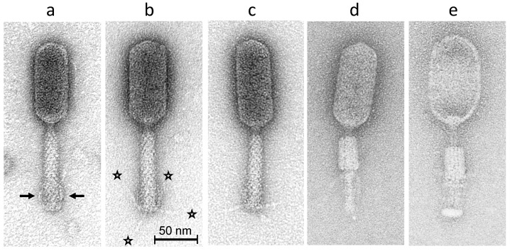
Figure 1.
Transmission electron micrographs of phage S144 negatively stained with 2% (w/v) uranyl acetate. Three intact phage particles with extended tail sheaths are shown in (a–c) with short and notably rigid tail fibers attached to the distal tail region in upward position (i.e., in retracted configuration indicated by arrows in (a) or in (randomly) extended configurations ((b,c); see asterisks in (b)). Phage particles with contracted tail sheaths are shown in (d) (with intact capsid) and in (e) (with empty capsid). Bar represents 50 nm, as indicated.

