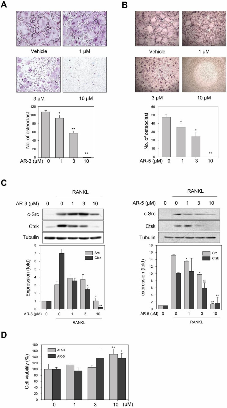
Figure 2.
AR-3 and AR-5 suppress RANKL-induced osteoclastogenesis in BMMs. (A,B) BMMs were treated with the indicated concentrations of AR-3 (A) or AR-5 (B), and then stimulated with M-CSF and RANKL for seven days. The cells were stained for TRAP. The number of TRAP-positive osteoclasts (>5 nuclei) was determined following image capture. Data are presented as the mean ± SE (* p < 0.05 and ** p < 0.01, versus vehicle-treated control; n = 3). (C) BMMs were treated with the indicated concentrations of AR-3 (left panel) or AR-5 (right panel), and then stimulated with RANKL for four days. Total lysates were prepared and the expression levels of c-Src and CtsK were determined by western blotting. Graphs represent densitometry analyses normalized to tubulin (* p < 0.05 and ** p < 0.01, versus vehicle-treated control; n = 3). (D) BMMs were incubated with M-CSF in the presence of the indicated concentrations of AR-3 or AR-5 for four days. Cell viability was measured using the MTT assay. Data are presented as the mean ± SE (* p < 0.05 and ** p < 0.01, versus vehicle-treated control; n = 3).

