Figure 1.
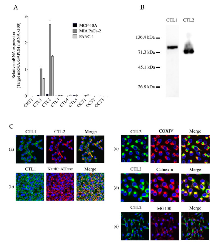
Expression of choline transporters. (A) Real-time PCR analysis of mRNA expression of CHT1, CTL1-5, and OCT1,2 in MCF-10A, MIA PaCa-2 and PANC-1 cells (n = 3). Relative mRNA expression expressed as ratio of target mRNA to glyceraldehyde-3-phosphate dehydrogenase (GAPDH) mRNA, which is a housekeeping gene. (B) Expression of CTL1 and CTL2 proteins in MIA PaCa-2 cells by Western blot analysis. (C) Intracellular distribution of CTL1 and CTL2 proteins in MIA PaCa-2 cells. (Ca) Subcellular distribution of CTL1 (green) and CTL2 (red) was determined by immunocytochemical staining. DAPI (blue) was used for nuclear staining in all specimens. Merged images labeled Merge, and yellow represents colocalization. (Cb) Subcellular distribution of CTL1 protein (green) analyzed using plasma-membrane marker Na+/K+-ATPase (red). CTL1 protein predominantly present on plasma membrane. Subcellular distribution of CTL2 protein (green) analyzed using mitochondria, endoplasmic reticulum (ER), and Golgi apparatus markers, (Cc) COX IV, (Cd) calnexin, and (Ce) MG130, respectively. CTL2 protein partially localized in mitochondria and ER but not colocalized in the Golgi apparatus.

