Figure 3.
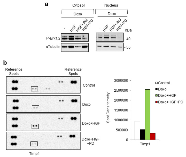
Cytokine profiles induced by HGF-Met-Erk1,2 preconditioning against Doxo damage. H9c2 cells were untreated (control) or treated with Doxo (25 μM), Doxo+HGF (0.5 nM), Doxo+HGF+PD (PD98059, 1 µM) (a,b) and Doxo+HGF+JNJ ((JNJ38877605, 500 nM) (a). Cells were treated with the inhibitors (PD and JNJ) for 4 h and exposed to Doxo in the last hour. For cell treatments, see Figure 1a. (a) P-Erk1,2 protein levels were detected in cytosol and nuclear fractions. αtubulin was used as the loading control. (b) Representative images (left) and densitometric analysis (right) of protein samples that were probed with the rat cytokine antibody array, that allowed analyzing 29 cytokines simultaneously. Data are representative results of three independent experimental replicates.

