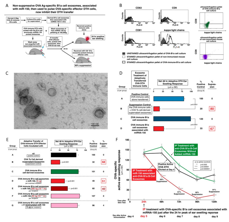
Figure 5.
Non-suppressive OVA Ag-specific B1a cell exosomes inhibit adoptively transferred OVA DTH when associated with miRNA-150. (A) Experimental protocol. (B) OVA-specific B1a cell-derived exosomes were cytometrically analyzed for expression of CD9, CD63, and CD81 as general markers of small exosomes, after coupling onto beads (left and center). Furthermore, they were analyzed and found positive for co-expression of CD9 and antibody kappa free light chains (FLC, center, and upper right), while some in the pellet from ultracentrifuged culture supernatant from normal lymphoid cells expressed Ab FLC, but did not co-express CD9 (right lower). (C) B1a cell-derived exosomes were analyzed with transmission electron microscope, showing the presence of bilamellar vesicles. (D) Non-suppressive OVA Ag-specific B1a cell-derived exosomes become suppressive of OVA DTH adoptive transfer when in vitro associated with miRNA-150 (Group C vs. D). (E) A dose response experiment shows that non-suppressive OVA Ag-specific B1a cell-derived exosomes are rendered suppressive of DTH effector cell adoptive transfer by their association in vitro with low doses of miRNA-150. (F) OVA Ag-specific B1a cell-derived exosomes associated in vitro with inhibitory miRNA-150 are able to suppress active OVA DTH when injected intraperitoneally (IP) at the 24-h peak of ear swelling response, in contrast to similar exosomes not associated with miRNA-150 (red line vs. green line, 50–92% suppression over the subsequent four days), and compared to no treatment of this active DTH (the thick black line). Results of in vivo assays are shown as delta ± standard error (SE), one-way ANOVA with post hoc RIR Tukey test. n = 5 mice in each group or three samples.

