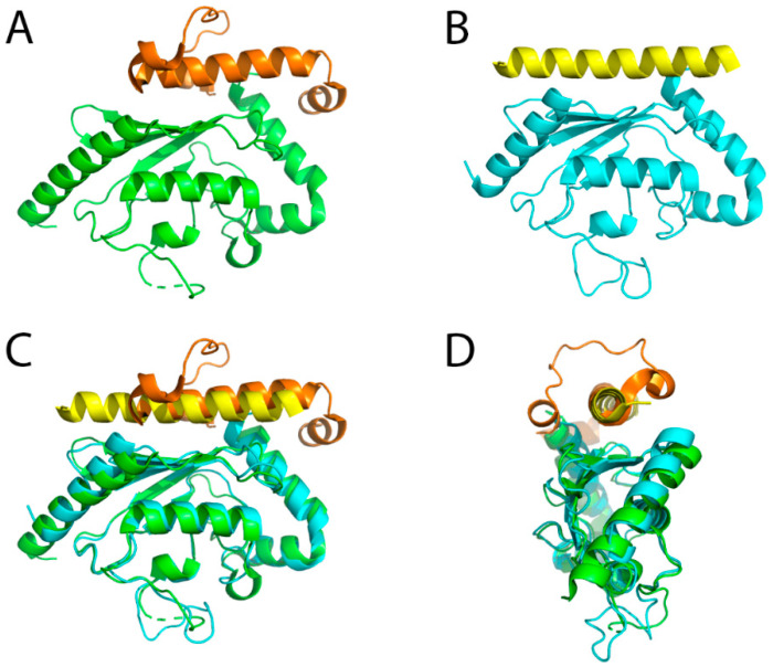
Figure 2.
Crystal structures of E2 enzymes in complex with the U7BR/G2BR domains. (A) Yeast Ubc7 in complex with U7BR (PDB ID: 4JQU). The green and orange colors represent Ubc7 and U7BR, respectively; (B) Mammalian Ube2g2 in complex with G2BR (PDB ID: 3H8K). The cyan and yellow colors represent Ube2g2 and G2BR, respectively; (C) Superimposed structures of (A,B); (D) Superimposed structures of (A,B), rotated 90° along the vertical axis compared to (C). In both cases the binding domain interacts with the backside of the E2 enzyme, opposite from the catalytic cysteine.

