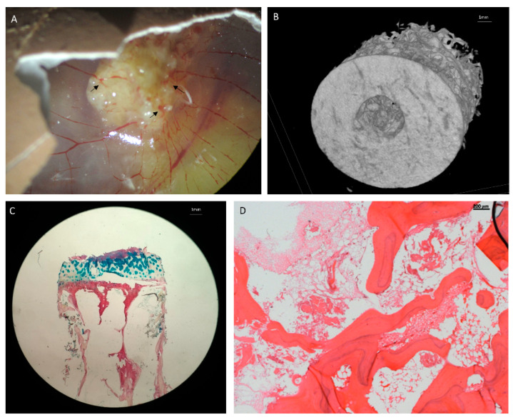
Figure 3.
Bone extracellular matrix (ECM) implanted in CAM model for vascularization. (A) Human bone inserted into CAM model. Arrows show areas of angiogenesis into the bone. (B) A µCT image of the bone model. (C) Alcian Blue and Sirius Red staining of the bone ECM, Blue denotes cartilage and proteoglycans, red denotes collagen. (D) Hematoxylin and Eosin Y staining of the bone ECM.

