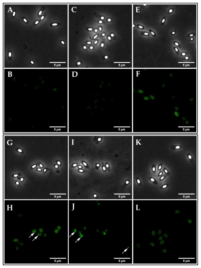
Figure 2.
Visualization of the GerRB-SGFP2 germinosome in B. cereus dormant spores. Phase-contrast (A,C,E,G,I,K) and fluorescence microscopy images (B,D,F,H,J,L) of dormant spores harboring various plasmids including: (A,B) wild-type spores; (C,D) spores carrying pHT315; (E,F) spores carrying pHT315-PaphA3′-SGFP2; (G,H) spores carrying pHT315-PgerR-gerRB-SGFP2; (I,J) spores carrying pHT315-PgerR-gerR-SGFP2; (K,L) spores carrying pHT315-gerRB-SGFP2 (no gerR promoter). Images were captured with a PH3 channel exposure time of 200 ms and an excitation channel at 470 nm using 10% laser power with an exposure time of 2 s The white arrows indicate some of the likely germinosomes in spores, and note the large heterogeneity in their fluorescence intensity. All panels are at the same magnification, and the scale bar is 5 μm.

