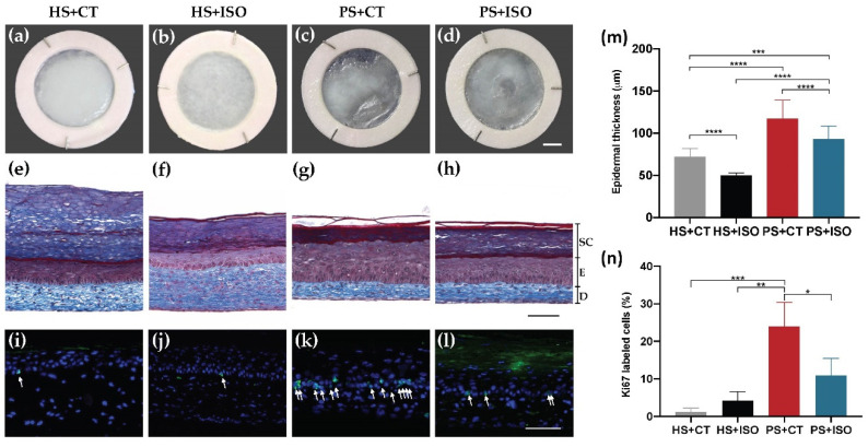
Figure 2.
Morphology and epidermal proliferation of healthy substitutes (HS) and psoriatic substitutes (PS) produced with cholera toxin (+CT) or isoproterenol (+ISO). (a–d) Skin substitute macroscopic aspects; (e–h) Masson’s trichrome staining of skin substitute histological sections (D: Dermis, E: Epidermis living layers, SC: Stratum corneum); (i–l) Ki67 immunofluorescence (green) detecting keratinocytes in proliferation. White arrows indicate Ki67-positive cells. Nuclei were stained with Hoechst (blue); (m) quantification of the thickness of the epidermal living layer (designated as E in panels (e–h) measured from Masson’s trichrome staining using the ImageJ software; (n) ratio of Ki67 positive cells to the number of total keratinocytes in the basal layer. Scale bars: (a–d) 5 mm; (e–h) 100 μm. Data presented are the means +SD (N = 2 donors per condition, n = 3 skin substitutes per donor). The statistical significance was determined using one-way ANOVA followed by a Tukey’s post-hoc test. (* p-value < 0.05; ** p-value < 0.01; *** p-value < 0.001; **** p-value < 0.0001).

