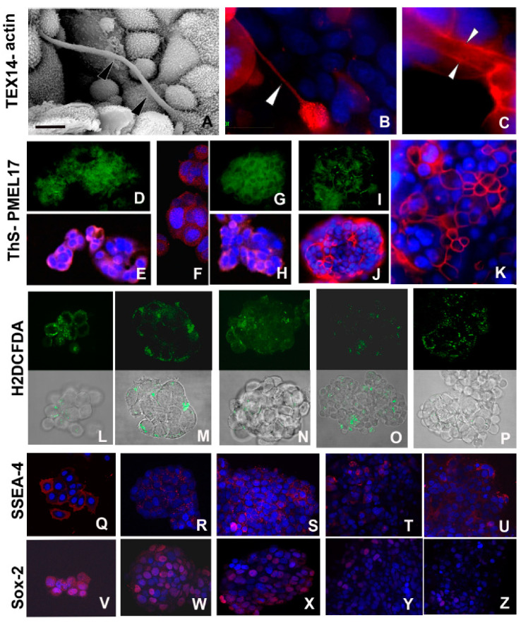
Figure 3.
Morpho-functional characterization of cells during early spheroid formation. (A–C) SEM (A) and immunofluorescence (B,C) analysis of MCF7 cells during the early phases of aggregation. Cell communications are established via TNTs, evidenced ultrastructurally (A) (arrowheads) and by immunofluorescence stainings (B,C). The nanocanals are confirmed by intensive labelling showing the expression of testis expressed gene 14 (TEX14) (B) (arrowhead) and of actin (C) confined along the internal face of TNT plasma membrane (arrowheads). Nuclei are stained with 4′,6′-diamino-2-phenylindole (DAPI) and marked in brilliant blue. Scale bar: (A) 7 μm. (D–K) Identification of amyloid fibrils localized by bright green fluorescence of thioflavine S (ThS) and by expression of melanocyte protein (Pmel17) (red) in MCF7 cells during early aggregation phase (24h) (D,E), after 3 days (F,H), and only superficially in mature spheroids at 5–7 days (I–K). Nuclei are stained with DAPI. Separate staining for ThS (D,G,I) and Pmel17 (E,F,H,J,K), are proposed to better evaluate each signal identifying the presence of amyloid materials. (L–P) The overall degree of cytoplasmic ROS is detected using the fluorescence dye 2′,7′-dichlorodihydrofluorescein diacetate (H2DCFDA). Comparison among the upper series of photographs showing only the staining for ROS with the related lower series, showing superimposed bright field images and ROS localization, is proposed to provide a better evaluation of the signal strength and its localization in the various developmental stages of spheroid. The signal (green) persists during all developmental stages of spheroid with not-significative decrease in the last phase of development. Early (24 h) aggregation phase (L,M), 3 days spheroid (N), 5–7 days (O), and 11–16 days spheroid culture (P). (Q–Z) Comparison of the expression of stemness markers stage-specific embryonic antigen-4 (SSEA-4) (Q–U) and SRY (sex determining region Y)-box 2 (Sox2) (V–Z) from 24 h aggregates (Q,V), and 3 (R,W), 5–7 (S,X), and 11–16 (T,Y,U,Z) days spheroids. The signal decreases along with maturation of spheroids and it is superficially confined, as evidenced in the overview of selected peripheral cells of spheroid 11–16 days old (U,Z)

